AI in Healthcare and Medicine

Back to Verticals page

ani-kolleshi-7jjnJ-QA9fY-unsplash


AI in health and medicine is a game-changer, revolutionizing diagnostics, treatment, and healthcare management with a focus on predictive analytics and personalized medicine, marking a transformative impact at the forefront of medical innovation.

a. Nucleus Detection and Segmentation

This project focuses on advancing computational pathology by tackling the complex task of nucleus detection in histology images. We propose an innovative approach that integrates spatially constrained context-aware correlation filters derived from hierarchical deep features. To further enhance the robustness and accuracy of our method, we employ a multi-level feature fusion strategy. This fusion technique is guided by inter-pool consistency and pool robustness, contributing to improved nucleus detection performance. A key aspect of our approach involves addressing challenges commonly encountered in histology images, such as nuclear clutter and irregular boundaries. To overcome these issues, we introduce structural graph-based constraints providing a more effective solution to the intricate task of nucleus detection.

Related Publications

b Nucleus Classification

This study focuses on advancing computational pathology for tumor micro- environment profiling by introducing novel approach to identify nuclear components in histology images. Unlike conventional methods, we employ a fully learnable message-passing network using a nearest-neighbor graph to capture physical interactions among nuclei. By computing appearance and geometric features, our algorithm efficiently diffuses contextual information, leading to improved nucleus classification performance. Our method predicts biologically meaningful nuclear communities, contributing to a more comprehensive understanding of the tumor microenvironment.

Related Publications

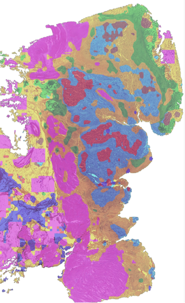
c. Tissue Phenotyping

This study will aim to advance tissue phenotyping in cancer histology images within computational pathology. In the first approach, tissue phenotypes are treated as communities within cellular graphs, utilizing a deep neural network for improved cell detection and classifican. This method enhances tissue phenotyping by constructing both cell-level and patch-level graphs. The second study introduces an innovative algorithm that outperforms existing methods by integrating cell-level features within a hierarchical graph-based framework. By leveraging texture, alpha diversity, and multi-resolution deep features, the algorithm constructs a multiplex cellular community-based network, contributing to more accurate and detailed tissue phenotyping.

Related Publications

d. Skin Cancer Segmentation

This work introduces an innovative and lightweight method for foot ulcer segmentation, setting it apart from established techniques in the field. The proposed model integrates residual connections, channel attention, and spatial attention within convolution blocks. Notably, our approach diverges from widely used backbone architectures, pre-training, and transfer learning strategies. The training methodology employed is a straightforward patch-based approach, enriched with test time augmentations and majority voting. This simplicity in training underscores the effectiveness of our lightweight solution, providing a compelling alternative to the more intricate architectures commonly utilized in foot ulcer segmentation tasks.

Related Publications

e. Prostate Cancer Detection

A method for Gleason score classification method is proposed in prostate tumors using MRI data. Patches extracted from annotated MRI images are used as features after encoding using a dictionary. Our voting-based encoding method transforms the data into more discriminative class-specific representations. In essence, our research introduces a novel methodology for Gleason score classification in prostate tumors.

Related Publications

f. ECG Estimation Using Low-Cost Portable PPG Sensors

The bio-markers produced by ECG under clinical settings are the foremost methodology in detecting Cardiovascular disorders. However, the ECG monitoring systems are costly and generally not portable. Therefore, in this project we propose utility of low cost and portable sensors such as Pulse Oximeters for estimation of ECG signal using Artificial Intelligence. Photoplethysmography (PPG) measures changes in blood volume and it is limited to the amount of information it yields to determine cardiovascular disorders. The introduction of a technique that accurately extracts the necessary information that exists in the ECG signal from the PPG signal would go a long way in making the process of cardiac disease detection easier, non-invasive and not requiring strict clinical settings. One potential application of PPG to ECG translation is in the monitoring of patients with heart conditions. For example, a person with atrial fibrillation (AFib) may not always experience symptoms, but their heart rhythm can still be irregular. By continuously monitoring their PPG and translating it to ECG, any changes in heart rhythm can be detected and addressed promptly. Another potential application is in the monitoring of athletes during exercise. By continuously monitoring their PPG and translating it to ECG, changes in heart rate and rhythm can be detected and used to adjust the intensity of the exercise. Overall, PPG to ECG translation has the potential to improve the accuracy and convenience of heart rate and rhythm monitoring. By using machine learning algorithms, PPG readings can be translated to ECG readings in real-time, allowing for continuous monitoring using only a pulse oximeter. This has the potential to improve the care of patients with heart conditions and to enhance the performance of athletes.

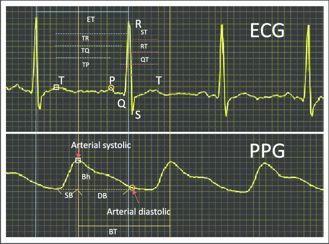
g. Emotion Detection Using ECG Signal

Emotion Detection Using ECG Signal involves analyzing the heart's electrical activity (ECG) to discern emotional states. By applying signal processing and machine learning to ECG data, this technology aims to identify patterns correlating with different emotions.

Related Publications

h. Classification of Benign and Malignant Masses in Mammograms

This study addresses the challenge of classifying benign and malignant masses in mammograms, a pivotal aspect in the development of Computer-Aided Diagnosis (CAD) systems for breast cancer detection. To overcome the complexities associated with mass region segmentation, our approach shifts the focus to the entire Region of Interest (RoI). We explore the application of the Bag of Visual Words (BoVW) technique, treating the RoI as a collection of local features. We investigate various local features and methods for constructing a code-book, followed by the implementation of a voting-based approach for feature encoding. This approach aims to enhance the robustness and accuracy of the classification process for distinguishing between benign and malignant masses in mammograms.

Related Publications

i. Unsupervised WSI Classification

This project aims to develop fully unsupervised algorithm for gigapixel Whole Slide Image (WSI) classification using mutual transformer learning, which transforms image patches into a latent space and generates pseudo-labels through iterative training. The framework showcases its effectiveness in unsupervised WSI classification and extends its capabilities to weak supervision for cancer subtype classification as a downstream analysis. By minimizing the reliance on manual annotations, the proposed approach offers a practical solution for gigapixel WSI classification without compromising performance in computational pathology applications.

Related Publications

● Unsupervised Mutual Transformer Learning for Multi-Gigapixel Whole Slide Image Classification, arXiv, 2023

● CPLIP: Zero-Shot Learning for Histopathology with Comprehensive Vision-Language Alignment, CVPR, 2024